Brain lessons
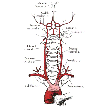
It's been a while since it occurred now, but I know lots of people are still interested in what caused Linda's stroke. I am far from a brain expert, but I can explain it as I understand it from the summaries I got from doctors. First it helps to understand something about how blood gets into the brain. Like all parts of the body, arteries bring oxygenated blood from the heart and veins bring blood back. In diagrams of the body, you'll often see arteries in red and veins in blue. The following diagram (stolen shamelessly from the internet) shows arteries only, but it has the detail needed to understand Linda's stroke.
My apologies in advance to anyone who really knows how this stuff works.
Most everyone is familiar that the large arteries on each side of your neck are the carotids. More important to Linda's stroke are the smaller ones in the back that run up each side of the neck vertebra. They are called the vertebral arteries. Notice that they join together at the base of the scull to form a singular artery called the basilar artery. This artery forms the primary blood supply for the part of the brain that was damaged in Linda's stroke, the Pons.
The following image is one of Linda's actual MRI films. You'll notice that the carotids show up very prominently, and behind them the vertebral arteries can be identified, joining to form the single basilar artery.
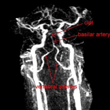
What is significant about the film is that the basilar artery just stops. There are not left and right branches extending up from it as you would expect after seeing the diagram above. As it was explained to me, an MRI shows flowing blood. If there isn't a flow, there isn't an image. So you don't see a clot as such in an MRI, but you detect its presence by the lack of flow where you expected to find it. Linda apparently had a clot in her basilar artery which interrupted the flow of blood to both the left and right side of her brain.
In the very plain words the neurologist used only minutes after seeing Linda, "This is bad." These kinds of strokes kill people. Obviously that didn't happen this time, although it has left us with a lot to recover from. Interestingly, the anatomy of the brain provides a mechanism for adapting to the clot. In the diagram, just above where the basilar artery branches, you'll notice that lots of arteries join together in a kind of circuit. This is referred to as the circle of Willis.
In a healthy brain, the typical blood flow would be up the basilar artery to the left and right branches and back through veins (not drawn) with some "leakage" into this circle. With the basilar blocked, you can expect these leaks to reverse direction and blood to arrive in the Pons from the circle instead. Now, you'll notice on the diagram that these vessels are drawn pretty small. This isn't the ideal way to get blood, but it can apparently keep you alive if you are lucky. Over time, the vessels can dilate and provide more blood, but it isn't immediate, and in the meantime brain tissue is going to be damaged and die. As we understand it, brain tissue does not regenerate. You have to learn to get by with what is left, and that is what rehabilitation is for. You retrain other (nearby) tissue to take over tasks that used to be done by the parts that were lost.
And of course our rehabilitation is going very well, which, in the end, is probably more important than exactly what caused the stroke.
It's been a while since it occurred now, but I know lots of people are still interested in what caused Linda's stroke. I am far from a brain expert, but I can explain it as I understand it from the summaries I got from doctors. First it helps to understand something about how blood gets into the brain. Like all parts of the body, arteries bring oxygenated blood from the heart and veins bring blood back. In diagrams of the body, you'll often see arteries in red and veins in blue. The following diagram (stolen shamelessly from the internet) shows arteries only, but it has the detail needed to understand Linda's stroke.
My apologies in advance to anyone who really knows how this stuff works.
Most everyone is familiar that the large arteries on each side of your neck are the carotids. More important to Linda's stroke are the smaller ones in the back that run up each side of the neck vertebra. They are called the vertebral arteries. Notice that they join together at the base of the scull to form a singular artery called the basilar artery. This artery forms the primary blood supply for the part of the brain that was damaged in Linda's stroke, the Pons.
The following image is one of Linda's actual MRI films. You'll notice that the carotids show up very prominently, and behind them the vertebral arteries can be identified, joining to form the single basilar artery.
What is significant about the film is that the basilar artery just stops. There are not left and right branches extending up from it as you would expect after seeing the diagram above. As it was explained to me, an MRI shows flowing blood. If there isn't a flow, there isn't an image. So you don't see a clot as such in an MRI, but you detect its presence by the lack of flow where you expected to find it. Linda apparently had a clot in her basilar artery which interrupted the flow of blood to both the left and right side of her brain.
In the very plain words the neurologist used only minutes after seeing Linda, "This is bad." These kinds of strokes kill people. Obviously that didn't happen this time, although it has left us with a lot to recover from. Interestingly, the anatomy of the brain provides a mechanism for adapting to the clot. In the diagram, just above where the basilar artery branches, you'll notice that lots of arteries join together in a kind of circuit. This is referred to as the circle of Willis.
In a healthy brain, the typical blood flow would be up the basilar artery to the left and right branches and back through veins (not drawn) with some "leakage" into this circle. With the basilar blocked, you can expect these leaks to reverse direction and blood to arrive in the Pons from the circle instead. Now, you'll notice on the diagram that these vessels are drawn pretty small. This isn't the ideal way to get blood, but it can apparently keep you alive if you are lucky. Over time, the vessels can dilate and provide more blood, but it isn't immediate, and in the meantime brain tissue is going to be damaged and die. As we understand it, brain tissue does not regenerate. You have to learn to get by with what is left, and that is what rehabilitation is for. You retrain other (nearby) tissue to take over tasks that used to be done by the parts that were lost.
And of course our rehabilitation is going very well, which, in the end, is probably more important than exactly what caused the stroke.
<< Home